
Our vascular services deals with the diagnosis, treatment and management of conditions affecting the arteries or veins and lymphatic vessels in the body, including:
- AAA REPAIR
- PERIPHERAL ARTERY BYPASS
- CAROTID DISEASE
- DIABETIC FOOT
- LEG ULCERS
- LYMPHOEDEMA & SWOLLEN LIMBS
- PERIPHERAL ARTERIAL DISEASE (PAD)
- VARICOSE VEINS
Abdominal aortic aneurysm (AAA) repair
The traditional operation involves making an incision into the abdomen to replace the aneurysm with an artificial piece of artery (a graft). This is a major operation and carries some risk. However, it is successful in most cases and the long-term outlook is good. In most cases, the graft will function for the rest of a patient’s life.

Complications: The risk of a major complication is about 5-6 % from an open operation. As with any major operation there is a small risk of you having a medical complication such as:
- Heart attack
- Stroke
- Kidney failure
- Chest problem
- Loss of circulation in the legs or bowel
- Infection in the graft used to replace the aorta
Up to 1 in 10 men may have difficulty obtaining an erection following surgery due to injury to nerves which lie on the front of the aorta.
Deep vein thrombosis (DVT) is a recognised risk and most patients will have treatment during their stay in hospital to prevent this. Patients developing a DVT may require extra treatment for this, which may prolong their stay in hospital.
If patients are deemed to be at higher risk of a major complication occurring, it is usually because there are already other existing medical issues. The surgeon will discuss this with the patient. It is important to remember that the surgeon will only recommend treatment for an aneurysm if he or she believes that the risk of the aneurysm bursting is higher than the threat posed by the operation. The surgeon will be able to outline the success rate for this operation in his/her unit.
Bypass of a Peripheral Artery
A bypass procedure is the commonest open surgical procedure carried out in the lower limb for ischaemia. The precise name given to the procedure depends on where the bypass starts and finishes. Therefore, the procedure could be termed as an aorto-femoral, ilio-femoral, femoro-popliteal, femoro-tibial or popliteal-pedal bypass. Artificial Dacron or Polytetrafluoroethylene (PTFE) grafts are used to bypass larger arteries. Below the groin the smaller femoral and popliteal vessels are often best bypassed using the patient’s own vein. The long saphenous vein is ideally situated for this.
These procedures require a general or regional anaesthetic. Therefore, the fitness of the patient is a key factor in deciding whether or not to expose them to the risks of surgery.
Pre-operative preparation is also vitally important in order to optimise the patient’s cardiac, respiratory, and renal function.
The principle underlying these bypass operations is to expose and control the inflow and outflow artery above and below the diseased segment. The arteries can then be clamped and opened and the graft sewn on at both ends. When the clamps are released, blood will then flow through the graft bypassing the diseased segment.
Major complications occur in approximately 5-8% of cases. The commonest local complications after surgery are thrombosis leading to occlusion of the graft, bleeding, and infection. The latter is more common with artificial grafts. Patients with vascular disease frequently have co-morbid conditions and are therefore, prone to a number of general postoperative problems. These can include heart attack, chest infection, deep veinous thrombosis / pulmonary embolism and reduction in kidney function. Good postoperative care helps to minimise these complications.
Surgery is usually reserved for severe limb ischaemia, where there is concern that leaving the poor circulation will damage the leg and risk amputation. The risks of surgery are then worthwhile.
Carotid Disease

The carotid arteries carry oxygenated blood to the head, brain and face. The common carotid artery runs up the neck where it divides into two smaller branches – the internal and external carotid arteries.
Carotid artery disease occurs when plaque (a waxy substance) builds up inside the carotid arteries. This build up, known as atherosclerosis, can result in symptoms of arm weakness, changes in facial muscles with a ‘droop’ on one side, and muddled or loss of speech. A temporary loss of vision in one eye, known as amaurosis fugax, is also a warning sign. These symptoms often resolve over 24 hours but may reoccur.
The symptoms listed above are commonly associated with what is known as a transient ischemic attack (TIA), and act as a warning sign that a more significant stroke may occur without treatment.
The carotid arteries in the neck can be investigated by undertaking an ultrasound, CT or MRI scan. These are non-invasive scans that can detect significant narrowing of the internal carotid artery.
If you experience a TIA and a scan indicates a significant narrowing of the carotid artery, you are at the risk of a major stroke. Studies have shown that the risk can be reduced by up to 6% following surgery and stenting. A surgical procedure known as a carotid endarterectomy is commonly performed to remove plaque build up from the carotid artery.
The risk factors for vascular disease, such as high blood pressure, cholesterol, stopping smoking, and diabetes should also be addressed.
Diabetic Foot
Over time, diabetes can lead to peripheral nerve damage, accelerated peripheral arterial disease and a reduced ability to fight infections. These factors can make the foot vulnerable, requiring extra care and vigilance. Annual checks often include an inspection of the feet by a trained healthcare professional.
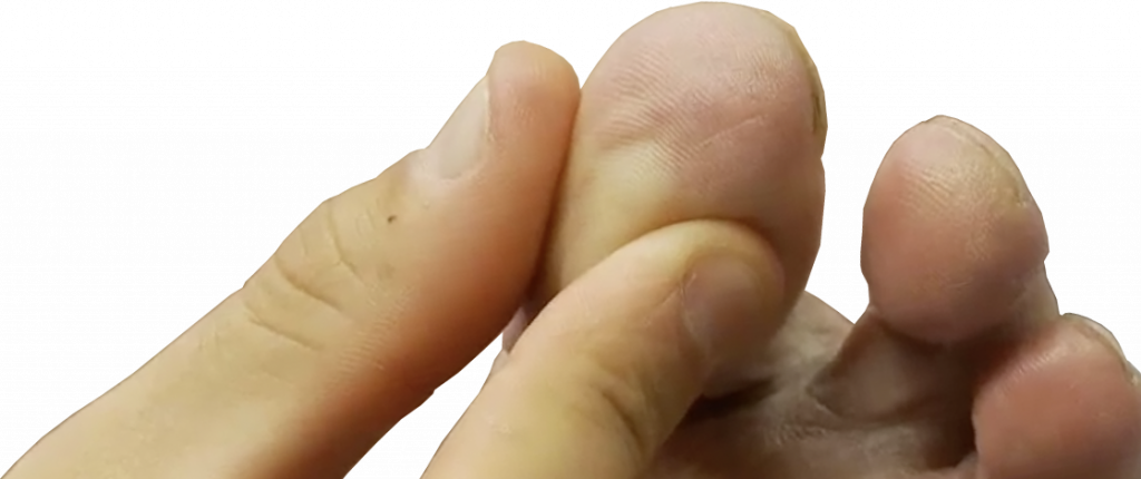
The nerve function and pulses in each foot are checked, as well as any noticeable changes in the shape of the foot. Footwear advice is provided, and a podiatrist may treat the feet if required. The management of diabetic control is also reviewed. These investigations are designed to identify and prevent problems occurring. If you are experiencing any of these symptoms, you will be monitored more closely and offered extra advice on foot care. This may include special footwear to protect your feet.
Leg Ulcers
What is a leg ulcer?
A leg ulcer is caused by a break in the skin on the leg that allows air and bacteria enter the underlying tissue, often caused by a minor skin injury.
In most people, such an injury will heal up within a week or two. However, when there is an underlying problem, the skin does not heal and the area of ulceration (breakdown) can increase in size. This is a chronic leg ulcer.
The most common underlying problem causing chronic leg ulcers is disease of the veins in the leg. Venous disease is the main reason for over two thirds of all leg ulcers.
- Venous Disease (caused by veins not working) – about 80% of leg ulcers
- Arterial Disease (caused by the arteries not working) – about 15% of leg ulcers
- Other causes (includes diabetes and rheumatoid arthritis as well as some rare conditions) – about 5% of leg ulcers
In some cases, you may experience two or more of these conditions simultaneously. Your doctor will examine you and perform some tests to determine what type of ulcer you have. The following advice applies to venous ulcers and may not be appropriate for other types of ulcers.
The veins in your leg carry the blood back from the foot towards your heart. They include one-way valves that ensure the blood flows in the correct direction. In some people, these valves are not very effective or can be damaged by thrombosis (clots). Damage to the valves can cause the blood to flow in the wrong direction, which results in high pressure within the veins when standing. This abnormally high pressure damages the skin and leads to ulcer formation.
How will it be treated?
Treatment of a venous leg ulcer can be approached by two methods:
- Controlling the high pressure in the leg veins
- Treatment of the ulcer
The mainstays of treatment are elevation of the leg, compression bandaging or stockings, and vein interventions.
Elevation of the leg
The higher the leg is elevated, the lower the pressure in the veins will be. If the foot is elevated above the heart, the pressure in the foot drops to a normal level. Elevate your legs whenever you can and as high as you are able. The arm of the sofa is good. Elevating the lower end of your bed by about six inches can also help during sleep. You can use some old books for this.

Compression bandaging or stockings
To help maintain a low blood pressure in the leg veins at the ankle when standing, you will be treated with compression bandages or stockings. Several layers of bandages may be required to obtain the necessary pressure. Once the ulcer has healed, compression stockings can prevent the ulcer from returning. These stockings are specially fitted and are much stronger than ordinary support tights. If you have difficulty putting on your stockings, you can buy a special stocking applicator.
Intervention to the Veins
A duplex scan allows the healthcare professional to assess the function of the veins. If there is a lot of incompetence in the surface veins of the leg (blood flows in the wrong direction), there is evidence that treating these veins can help with ulcer healing whilst preventing recurrence. Common procedures for the veins are endovenous ablation using heat (laser ablation or radiofrequency ablation) or chemical (foam sclerotherapy) injury to cause the faulty vein to scar up and block.
Dressings
The nurse will apply a number of different dressings under the bandages depending on the state of the ulcer itself. These dressings may well change as the ulcer progresses.
How long will it take the venous ulcer to heal?
Most ulcers will heal in three to four months, with some taking up to six months. A small proportion of ulcers will resist treatment and may not heal, but generally treatments are successful.
Lymphoedema and Swollen Limbs
Swelling of the foot and/or leg can have a number of causes. If both legs are affected it may suggest a more generalised cause such as fluid retention in the body. Kidney, liver and heart function can influence this. It may also be a side effect of medications. A long period of immobility with the legs dependent (below heart level) can lead to a build up of fluid, since we rely on the movement of the muscles in the leg to move the blood and fluid up out of the legs towards the heart. In some illnesses the protein level in the bloodstream drops and this causes fluid to move into the tissues, causing bilateral limb swelling. In some cases abnormal fat distribution leads to the appearance of limb swelling, which is mistaken for fluid.
When one limb is affected, it can point to a more local problem in the leg being the cause of the swelling. There are a number of possibilities:
- Venous insufficiency – poor return of fluid in the veins from the leg. (valve failure, DVT, varicose veins)
- Infection – tissues are inflamed and accumulate fluid which leaks from the circulation.
- Injury – after any injury the healing response involves some element of swelling
- Lymphatic failure – damaged, blocked, or absent lymph channels and glands.
Investigations are therefore required to establish which of the above problems is causing the swelling. From the vascular perspective, it is important to establish the function of the veins. Lymphoedema is different from venous oedema, and it may be clear that the swelling is lymphoedema from examination. Sometimes specific tests are required to obtain a more cohesive diagnosis.
Treatment
Treatment will be directed to the underlying cause. Compression with support stockings is often recommended, and vein treatments may also be useful. Difficult cases of lymphoedema may require more specialised therapy.
Lymphoedema
What is lymphoedema?
We all have a small amount of fluid (lymph) in our body tissues. This fluid leaves our blood system to provide water and nourishment to the tissues. Most of this fluid is collected by a system of drainage tubes, similar to blood vessels, called the lymphatic system. Lymphoedema is swelling caused by a build up of lymph in the limbs as a result of poor drainage.
Initially, the swelling is often noticeable at the end of the day and subsides at night. However, unless the swelling is treated properly, the fluid becomes fixed in the leg permanently.
What causes lymphoedema?
A lack of lymphatics is the most common cause for lymphoedema. If there are very few lymphatics, the swelling may begin from teenage years or sometimes earlier. This type of lymphoedema is called Milroy’s Disease. One leg is often worse than the other and sometimes only one leg is affected.
In less severe cases the lymphatics may be able to cope initially and only start to fail in old age. This is referred to as Lymphoedema Tarda.
Are there any other causes?
Surgery, radiotherapy or injury to the arm and leg can be another cause of lymphedema, which can damage the lymphatics of those structures. Damage resulting from radiotherapy is unavoidable if the cancer is to be cured.
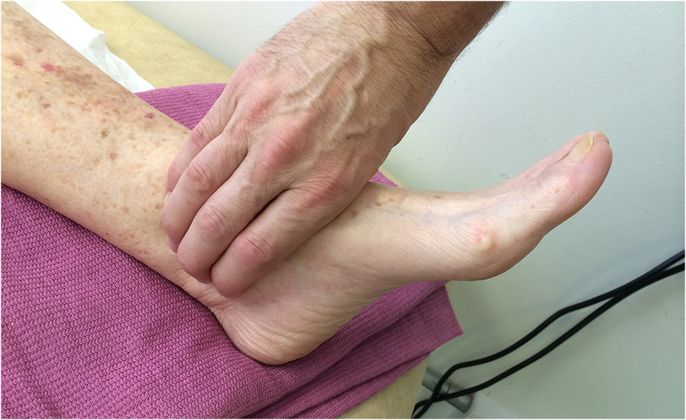
Peripheral Arterial Disease (PAD)
Narrowing of the arteries is referred to as peripheral arterial disease. It’s a common problem that affects 9% of the population, but will only result in symptoms for a quarter of those people.
Arteries carry blood away from the heart and supply essential nutrients and oxygen to every part of the body. A normal artery is hollow with a smooth internal surface for the blood to travel over. Blood travelling through the arteries can sometimes leave behind deposits, known as plaque, which stick to the inside of the artery. This process is known as atherosclerosis. Gradually, over many years, these deposits can build-up and cause the inside of the artery to narrow. This is often referred to as ‘furring up’ of the arteries, though healthcare professionals call this ‘occlusive disease’.
PAD is occlusive disease that occurs in the peripheral or outer arteries of the body, such as the legs. However, if you have been diagnosed with PAD, it is likely that the arteries supplying your brain and heart will also be affected, leaving you at higher risk of suffering a heart attack or stroke.
The causes of peripheral arterial disease are:
- Smoking
- Diabetes
- Hypertension
- High cholesterol
- Diet and weight
Vascular disease can also be a hereditary condition. If you have a history of vascular disease in your family, you may want to discuss this with your healthcare practitioner.
Varicose Veins
Varicose veins are swollen and enlarged veins, usually usually appearing on the legs and feet. They often appear as blue or dark purple, with a lumpy, bulging or twisted appearance.
The veins in the body rely on a series of valves to keep the blood flowing in the correct direction. If these valves do not function correctly, the blood flow slows or is reversed. There are several methods for treating varicose veins, which are outlined below.
If the function of your veins needs to be assessed, you will be asked to attend a special type of ultrasound scan, known as a Duplex.
What problems do varicose veins cause?
A build up of pressure in the veins can lead to pain and swelling. This may sometimes result in skin changes, such as brown staining, eczema or ulceration. A clot (thrombosis) may develop, causing the vein to become red, hard and tender. This is known as phlebitis.
There are several factors that can increase the risk of developing varicose veins:
- being female
- having a close family member with varicose veins
- being older
- being overweight
- having a job that involves long periods of standing
- being pregnant
- other conditions

Treatment options for varicose veins
Foam Sclerotherapy
This treatment is used to treat damaged valves and varicosities, and involves injecting chemical agents mixed with air to create a foam. The foam is injected into the veins, causing them to become inflamed. Scarring then occurs leading to obliteration of the vein. Ultrasound is used to direct the foam into the correct vein. The procedure can be performed in a clinic without the need for anaesthesia. Following treatment, patients are asked to wear a bandage or support stocking on the leg for two weeks.
The success rate for foam sclerotherapy is approximately 70 – 80%. If the veins do not disappear, treatment can be repeated. Some patients may experience side effects due to reactions resulting from the foam entering the general circulation. These reactions are uncommon and short-lived. Local phlebitis (inflamed thrombosis) of the treated vein can cause pain for 1-2 weeks and may occasionally result in scarring on the leg, though this only happens in 10 – 15% of cases.
Mechanico-chemical vein ablation
Performed under local anaesthetic, a sclerosant chemical is injected directly into the faulty saphenous vein trunk with a catheter. The catheter spins and modifies the lining of the vein, increasing the success rate for obliterating the vein. The long-term durability of this type of treatment is still being evaluated.
Endothermal Ablation
EndoVenous Laser Ablation (EVLA) and Radiofrequency Ablation (RFA) are alternative techniques for treating varicose veins. These procedures seal off the long saphenous vein (LSV) in the thigh, or the short saphenous vein (SSV) behind the knee and calf. Traditionally, the process was fairly invasive and involved tying and stripping the LSV and SSV. More modern ablation techniques have replaced surgery offering a less painful outcome with a shorter post-operative recovery period.
Both EVLA and RFA treatments can be performed under local anaesthesia. Using an ultrasound to aid guidance, a catheter is passed up the vein from the ankle or knee level and placed at the junction between the LSV / SSV and the deeper veins. When the catheter is activated, an electrical current or laser is passed through the vein wall, causing the proteins in the vein wall to change shape and contract down. As the catheter is slowly advanced down, the vein contracts until the blood flow has been closed. This method effectively removes the vein from the circulation, achieving the same outcome of the more traditional methods. Long-term follow up has shown that veins largely remain closed off and rarely open up again.
Phlebectomy
A series of tiny incisions are made in the skin through which a varicose vein is removed. This procedure is known either as an avulsion or phlebectomy. The veins are removed in sections through each of the incisions, which range from 2 – 4mm. Phlebectomy may be undertaken in conjunction with another form of varicose vein treatment, such as ligation and stripping.
After Care
The majority of these procedures are undertaken as day case or clinic treatments. An overnight stay in hospital following these treatment options is rare. The treated leg will be firmly bandaged to reduce bruising and assist with healing. After 1-2 days the bandage is often replaced with a stocking, which is worn for a further 5-7 days. It is often recommended to to wear the stocking during both the day and at night. Comfort may improve after a few days, after which point it is advised to wear the stocking during the day only. Patients are advised to either rest with the leg elevated whilst moving the ankle, or maintain their mobility around the house and garden. Standing for long periods of time and crossing legs should be avoided. It is suggested that patients undertake a daily walk of 1-2 miles to aid recovery.
Following ablation procedures, the treated vein may become sore after 3-5 days. This is a normal reaction to the treatment and will settle. Anti-inflammatory painkillers can help with initial discomfort. The small cuts from phlebectomy are often closed with skin glue or tape rather than stitches, though any stitches used are usually dissolvable.
Complications
Recovery from RFA or EVLA alone, without any phlebectomies, can last 2-4 days. If RFA and EVLA treatment also includes phlebectomies, this will inevitably produce some bruising and soreness. The severity depends on how many veins are removed, though the majority of soreness will settle in 3-5 days. Full recovery of bruising will take longer. Small nerves next to the veins can be disturbed, leading to patches of numbness in the lower leg and foot in approximately 10% of patients. This slowly resolves but is occasionally permanent. Less than 1% of patients may experience deep vein thrombosis (DVT) in the deeper veins of the leg. Maintaining mobility after the procedure and wearing a stocking will help reduce the risk of DVT. Infections in the very small wounds are uncommon.
Our Vascular Team
We have an expert team of vascular specialists, including consultants, specialist nurses, radiologists and anaesthetists involved in all aspects of care and treatment, from screening to surgery.
AAA Screening
Remember we offer Abdominal Aortic Aneurysm (AAA) screening to all men aged 65 and over for the Black Country. For further information please
Pre-operative Assessment
Pre-operative assessment is offered for all elective and urgent vascular patients mainly going through major vascular surgery. This includes aortic, carotid and peripheral arterial bypass surgery.